Beyond the Naked Eye: How New Dental Imaging Systems Are Changing Diagnosis
12/01/2026 02:09
30/03/2026 10:23
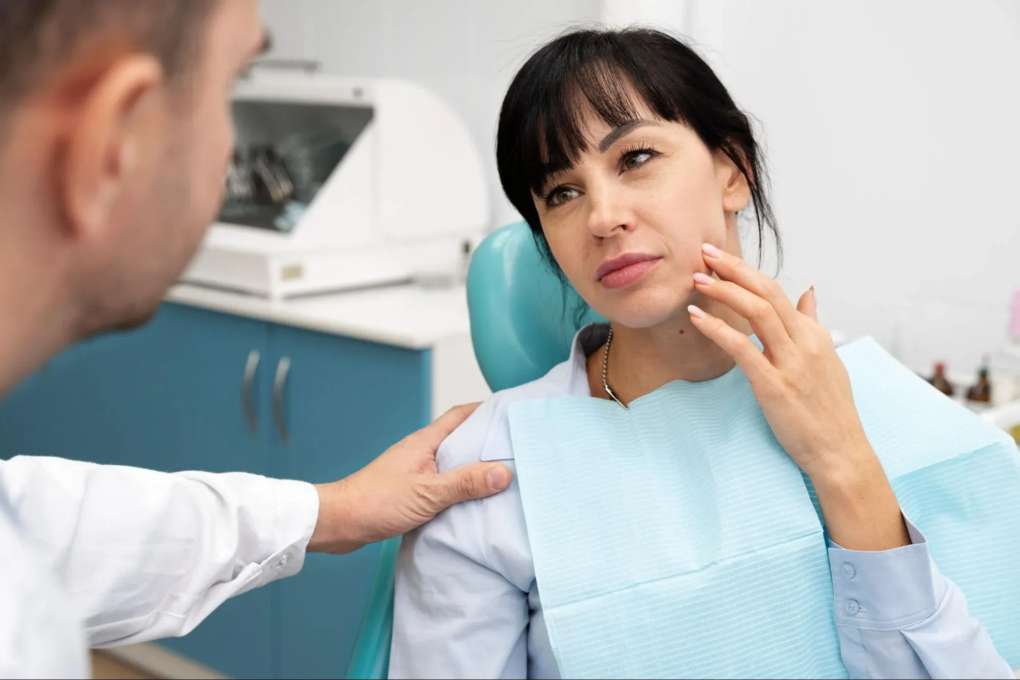
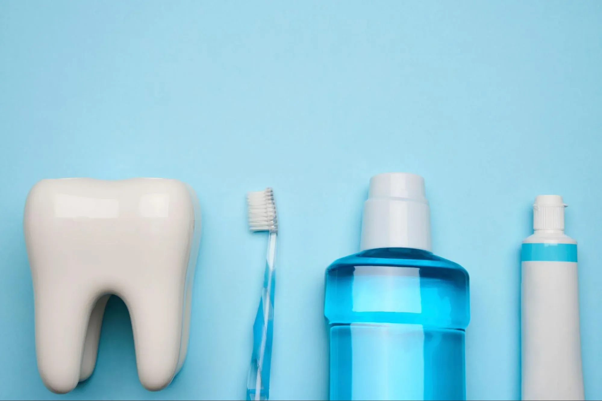
Healthcare moves quickly, and technology helps level the playing field. Like MRI changed brain surgery, new imaging systems have transformed dentistry. Dentists no longer rely only on mirrors, probes, and unclear X-rays to figure out what’s happening under your gums.
Today, leading dental hospitals work like digital command centers. Instead of just looking at teeth, we use advanced imaging tools to study them in detail. These systems help us see different layers, view the bones of the face, and spot problems when they first start.
This approach is called Advanced Dental Imaging. It’s not just one machine, but a group of tools that work together. For patients, this means three main benefits: safety with less radiation, faster diagnosis, and more accurate treatment planning.
If you’re a parent thinking about braces for your child or an adult planning implants, knowing about these tools helps you see the quality of care you’ll get. This guide will walk you through today’s dental imaging, including Cephalometric X-rays, RVG sensors, and how Artificial Intelligence is starting to help with diagnosis.
The Panoramic Evolution: The Digital Landscape
The Digital Panoramic X-Ray (OPG) is still the main tool for dental diagnosis. Panoramic imaging has been around for years, but new digital systems have made it much better.
-
Dynamic Focus: Old machines had a fixed focal point. If the patient moved slightly, the image blurred. Modern systems use "multilayer" technology, capturing multiple layers of focus simultaneously and automatically selecting the sharpest one.
-
Pediatric Modes: Advanced systems automatically reduce the radiation area for children, so they get only the smallest dose needed to see their growing teeth.
-
TMJ Views: These machines can take special pictures of the jaw joint with the mouth open and closed. This helps diagnose clicking or locking problems without needing a CT scan.
The Orthodontic Necessity: Cephalometric Imaging
If you or your child needs braces, you will meet the Cephalometric X-Ray (Ceph). Unlike a panoramic X-ray, which shows the teeth in a curve, a Ceph is a standardized side-profile view of the entire skull.
Why is this important? Orthodontics is about more than straightening teeth—it’s also about making sure the jaws are lined up correctly.
-
Skeletal Analysis: The Ceph lets the orthodontist measure how the upper and lower jaws relate to the base of the skull. For example, is an underbite because the lower jaw is too big or the upper jaw is too small? This answer changes the whole treatment plan.
-
Growth Prediction: By looking at the neck bones seen in the scan, doctors can tell a child’s “skeletal age” and predict how many growth spurts are left. This timing is important for planning orthopedic devices.
-
Airway Analysis: Advanced Cephs can measure airway width, helping diagnose whether a narrow jaw is causing sleep apnea or snoring.
RVG Sensors: The End of Film
For close-up pictures of single teeth, like checking for cavities or root canal infections, we use RadioVisioGraphy (RVG). These are small, wired sensors that go inside the mouth and replace the old, sharp plastic film.
-
Instant Results: There’s no waiting for film to develop. The image shows up on the computer as soon as the X-ray is taken.
-
Radiation Reduction: RVG sensors are the most sensitive X-ray detectors in medicine. They require 80% to 90% less radiation than traditional D-speed film.
-
Image Enhancement: Once the image is on the screen, the dentist can use filters. We can switch colors to see nerves better or use relief mode to make cracks stand out. This helps avoid mistakes.
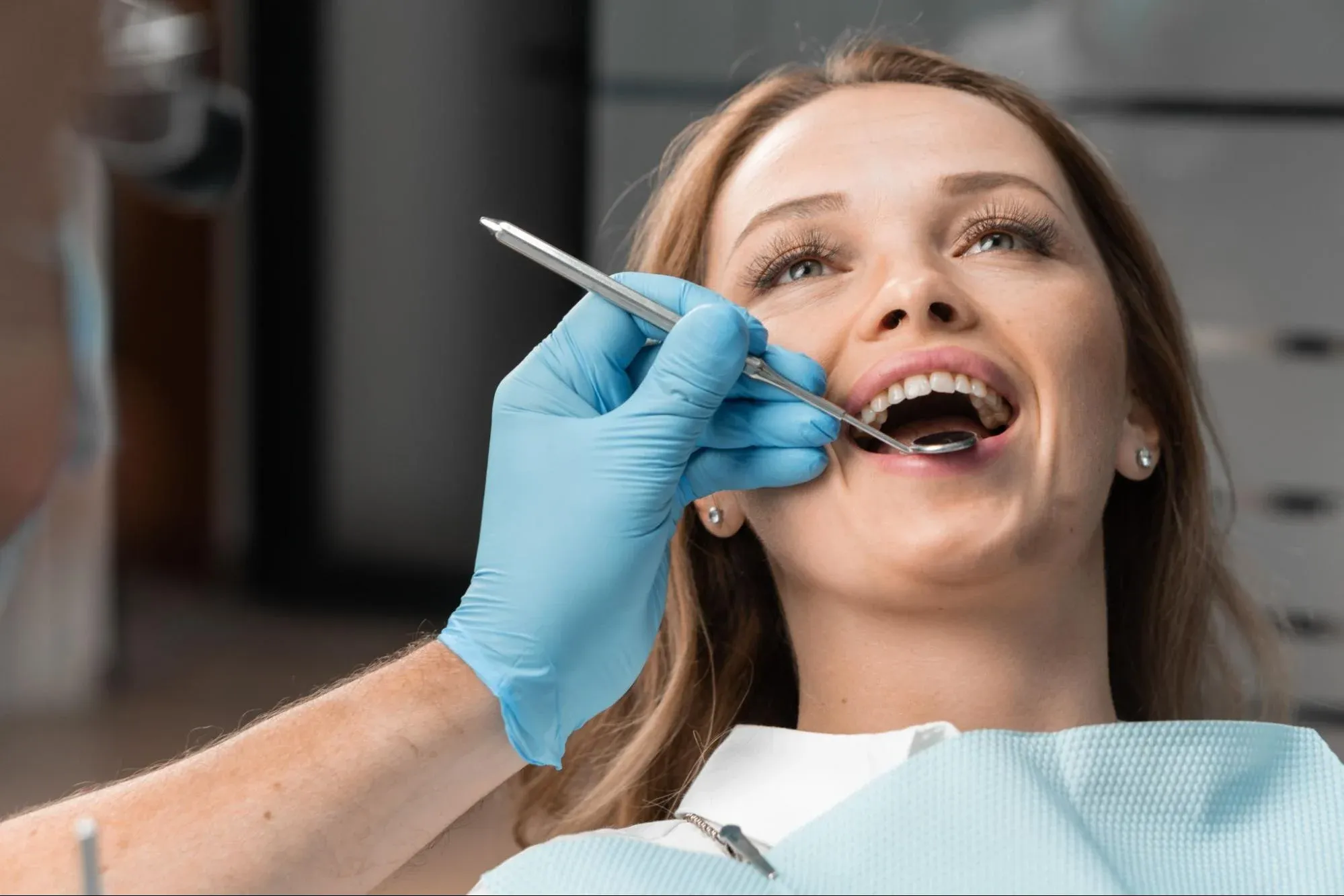
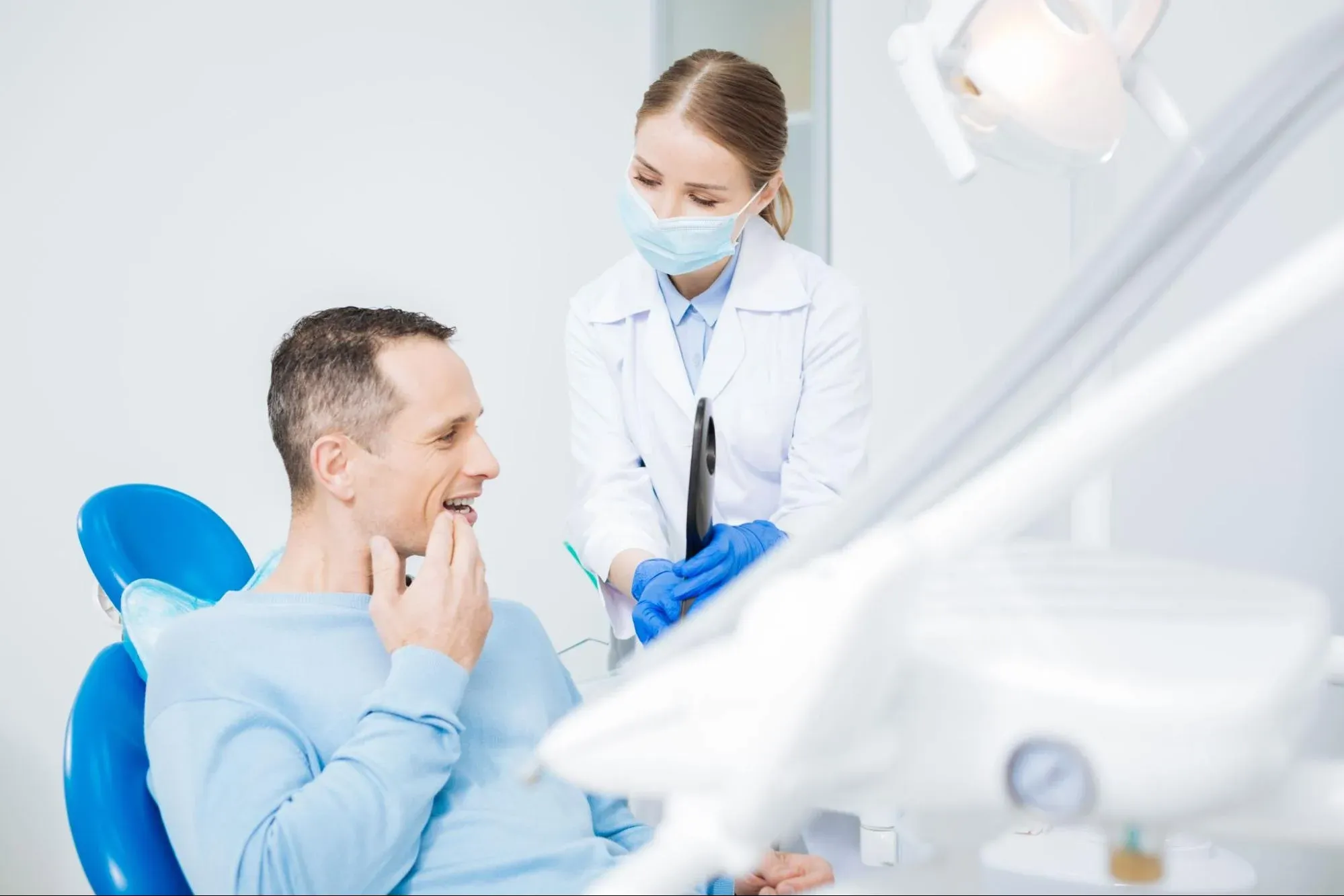
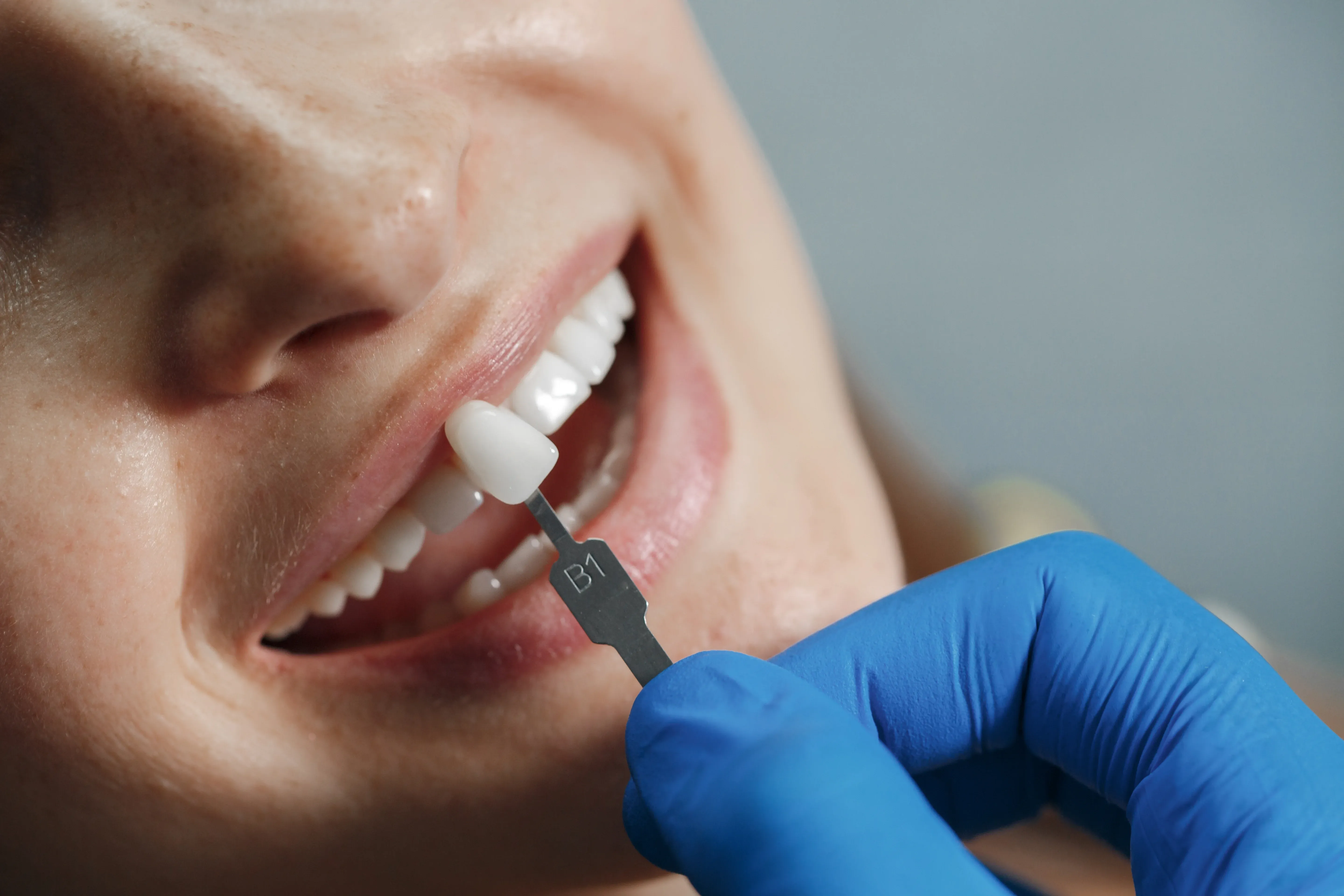
Intraoral Cameras: Seeing What the Dentist Sees
X-rays show the bone; Intraoral Cameras show the surface. These wand-like devices are high-definition video cameras with powerful LED ring lights.
-
The Patient Experience: Hearing “you have a cracked filling” can feel vague. But seeing your own tooth on a big screen, magnified 40 times, makes it clear.
-
Documentation: These images are a permanent record. We can compare your teeth from 2023 to 2025 to see if grinding is getting worse.
-
Hidden Details: The camera’s close-up lens can find tiny cracks or leaks in old crowns that we can’t see with the naked eye.
Artificial Intelligence (AI) in Diagnostics
The newest frontier in advanced imaging is the integration of AI Software. When an X-ray is taken, AI algorithms scan the image in the background.
-
Caries Detection: The AI works like a second set of eyes, marking spots between teeth that could be early signs of decay.
-
Bone Loss Measurement: AI can automatically measure how much bone is lost around each tooth, giving an objective score for gum disease severity.
-
Pathology Alerts: AI can spot rare issues like cysts that a person might miss while looking at the teeth. AI doesn’t replace the dentist, but it helps them be more accurate and lowers the risk of mistakes.
Phosphor Plate Systems (PSP)
For people with small mouths or a strong gag reflex, the hard RVG sensors can be uncomfortable. That’s why advanced clinics use Phosphor Plate (PSP) systems. These are thin and flexible, so they fit the mouth better. After use, they go into a laser scanner to make a digital image. PSPs are as comfortable as film but have all the benefits of digital technology.
The Safety Shield: Dose Management
Advanced imaging isn’t just about clearer pictures—it’s also about keeping patients safe. Modern machines have Automatic Exposure Control (AEC). They do a quick scan to check bone density. For a small child or petite adult, the machine lowers the X-ray power. For a larger person, it increases it a bit. This way, everyone gets just the right amount of radiation for a good image.
Integrated Workflows: The Digital Thread
The real strength of these systems is how they work together. In modern hospitals, Cephalometric tracings, 3D bone scans, and Intraoral Camera images are all combined in one software. This creates a Virtual Patient. We can plan surgery on the 3D scan, design a smile on the photo, and put them together. This smooth process from diagnosis to treatment is what sets today’s dentistry apart.
Diagnosing with Confidence
Ultimately, advanced imaging systems are tools of trust. They remove the ambiguity from healthcare. They allow the dentist to say, "I know," rather than "I think."
For patients, this means fewer surprises. Implants can be placed safely away from nerves. Braces can align the whole face, not just the teeth. Cavities can be found when they’re tiny and easy to fix.
At İstinye University Dental Hospital, our Radiology Department is at the center of what we do. We use the latest imaging systems, from low-dose Cephalometrics to AI-assisted analysis, because we believe great treatment starts with great vision. We look for what can’t be seen, so we can protect your smile.