Seeing the Invisible: How Micro-Endodontics is Revolutionizing Tooth Salvation
12/01/2026 00:45
30/03/2026 10:42
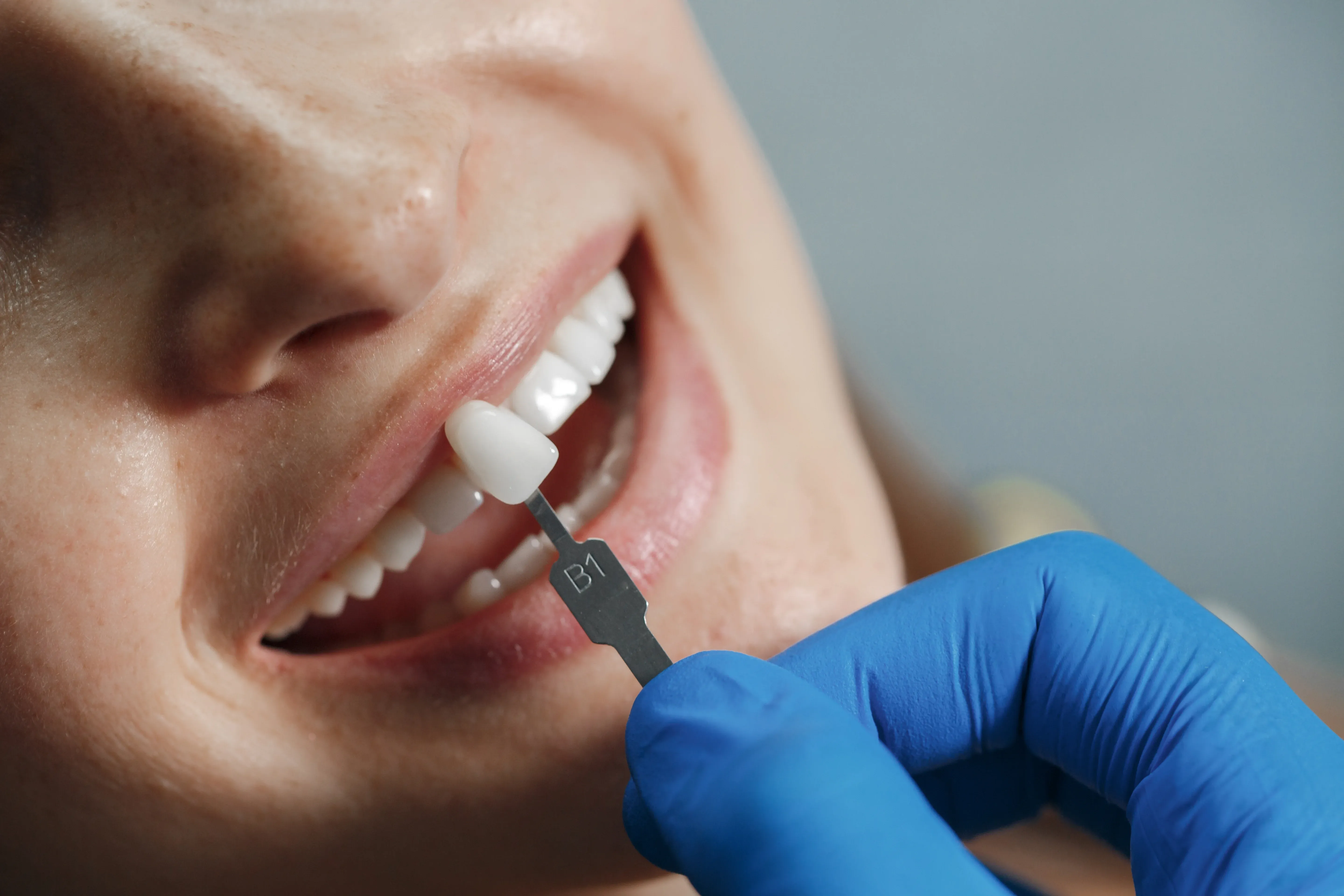
Dentistry requires great precision, but endodontics (root canal therapy) demands even more accuracy. The root canal system inside a tooth is a tiny maze, not just a straight tunnel. It has many small branches, twists, and hidden spaces.
For many years, dentists used their sense of touch and experience to work inside these small spaces. Even with magnifying glasses, many details of the tooth stayed hidden. Often, root canals failed not because of poor skill, but because dentists simply could not see everything they needed to.
The Dental Operating Microscope (DOM) has changed how dentists save teeth. This tool started a new approach called Micro-Endodontics. With strong light and up to 25x magnification, the microscope lets specialists see details they could not before. Now, instead of relying only on touch, dentists can actually see what they are doing, which means more teeth can be saved.
The Limitation of the Naked Eye
To see why this technology matters, consider how small the challenge is. The opening to a root canal is often tinier than a pinhead. Inside, the canal can split into two thin channels, curve sharply, or branch out like a river.
When a dentist works without magnification, they are essentially operating in the dark. They might miss a tiny fourth canal in a molar (a common cause of failure). They might miss a microscopic crack in the tooth's root. They might leave behind a pocket of infected tissue simply because it was tucked away in a shadow. The dental microscope eliminates this guesswork. It floods the inside of the tooth with coaxial light (light that travels the same path as the vision), ensuring there are no shadows, and magnifies the view so that a tiny canal looks as large as a highway.
The "MB2" Mystery: Finding Hidden Canals
One of the most compelling arguments for microscope-assisted endodontics is the infamous "MB2" canal. Upper molar teeth typically have three primary roots and three canals. However, studies have shown that in a significant percentage of people (60-70%), the first molar has a second canal in the mesio-buccal root, known as the MB2 canal.
This canal is very hard to find. It is often hidden by a layer of dentin and cannot be seen without special tools. If a dentist misses the MB2 during a root canal, bacteria can stay inside. The patient may feel fine at first, but the infection will come back and the root canal will fail.
With a dental microscope, the specialist can zoom in on the floor of the pulp chamber. They can distinguish the subtle color shift in the dentin that signals the entrance to this hidden canal by finding and treating the MB2, and the tooth's long-term prognosis skyrockets.
Treating Calcified Canals
As we get older, or if a tooth is injured or badly decayed, the tooth adds more dentin to protect itself. This can make the root canals shrink and become very narrow, sometimes closing up completely. To the naked eye, a calcified canal looks solid, with no opening.
Attempting to drill into a calcified tooth without magnification is risky. There is a great danger of "perforation"—drilling off-course and punching a hole through the side of the root, which can lead to tooth loss.
With the microscope, the dentist can see that calcified tissue looks a bit different from healthy tooth structure. They can carefully remove the calcification using special tools to find the original canal. This lets endodontists save teeth that might otherwise be considered untreatable.
Retreatment and Removal of Broken Instruments
Sometimes, a root canal has to be done again. This is called endodontic retreatment. It is needed if the first treatment did not heal properly or if a new infection appears. Retreatment is harder because the tooth now has old filling material, posts, or crowns inside.
Furthermore, a feared complication of root canal therapy is instrument separation. The tiny metal files used to clean the canals are fragile and can sometimes snap off, leaving a piece of metal wedged deep inside the root.
Removing a broken file without a microscope is like trying to retrieve a needle from a haystack while wearing boxing gloves. It often results in the removal of too much healthy tooth structure. With a microscope, the specialist can see precisely where the fragment is lodged. Using ultrasonic vibration, they can gently loosen and float the fragment out, clearing the path for proper cleaning without compromising the tooth's strength.
Detecting Micro-Fractures
Diagnosis is just as important as treatment. Sometimes, a patient has pain when biting, but there is no cavity and the X-ray looks normal. This is a sign of "Cracked Tooth Syndrome."
Vertical root fractures or tiny cracks in the crown often do not show up on X-rays. But with 20x magnification, these cracks are easy to see.
-
This matters because if a crack goes down into the root, the tooth usually cannot be saved. Root canal treatment will not fix it, since the crack lets bacteria in.
-
The benefit is that the microscope helps dentists make an early and clear diagnosis. This means patients do not have to go through a root canal on a tooth that cannot be saved. They can move straight to an implant or another solution, saving time and money.
Preservation of Tooth Structure: Minimal Intervention
In the past, dentists had to drill large openings to see inside the canals, which meant removing a lot of the tooth's crown. This made it easier to work, but it also weakened the tooth and made it more likely to break later.
Micro-endodontics follows the idea of Minimally Invasive Dentistry. The microscope gives such a clear view that the dentist can make a much smaller hole and only remove what is needed. By keeping more of the natural tooth, the tooth stays strong and can last for many years.
Apicoectomy: Microsurgery
Sometimes, a regular root canal cannot heal an infection at the tip of the root. In these cases, a surgery called an Apicoectomy is needed. The surgeon lifts the gum, removes the infected root tip, and seals it from the outside. This surgery requires a microscope, so the surgeon can check for cracks and seal the root with great accuracy.
The Patient Experience: Is it Different?
For patients, a procedure done with a microscope feels much like a regular one, but it is often more comfortable.
-
Ergonomics: The dentist sits upright and looks through the microscope, instead of bending over the patient. This relaxed position often leads to steadier hands and smoother work.
-
Communication: Many microscopes have video cameras that show images on a screen. The dentist can take a picture of a crack or hidden canal and show it to the patient. This helps patients understand what is happening in their mouth.
-
Speed: While setting up the microscope takes a moment, the procedure itself is often more efficient because the dentist isn't "hunting" for canals—they can see them immediately.
A Mark of Excellence
Buying a dental operating microscope is a big investment for any clinic, both in money and training. Having one shows that the clinic is committed to the highest standards of care. It means the dental team wants to do the best work possible, not just settle for "good enough."
Not every simple root canal needs a microscope, but having this technology for complex cases like curved roots, calcifications, or retreatments gives patients the best chance for success.
Saving a natural tooth requires precision. Moving from touch-based dentistry to vision-based micro-endodontics is the biggest advance in the field in the past 20 years. If you want to keep your natural smile despite infection or complex tooth anatomy, İstinye University Dental Hospital offers top-level endodontic care. Our specialists use advanced microscopes to find and treat even the most difficult cases, giving you the best chance for long-term success.