Seeing the Invisible: How Dental Radiology and Diagnostics Shape Your Health
12/01/2026 02:05
30/03/2026 10:19
When you smile in the mirror, you only see part of the story. About 60% of each tooth is visible above the gums, while the other 40%—including the roots, nerves, and jawbone—remains hidden. You also can't see where teeth touch, the health of your sinuses, or how dense your bone is.
In dentistry, treating a patient without seeing these hidden areas is like a pilot landing in heavy fog without instruments. It becomes guesswork, and when it comes to your health, that's not acceptable.
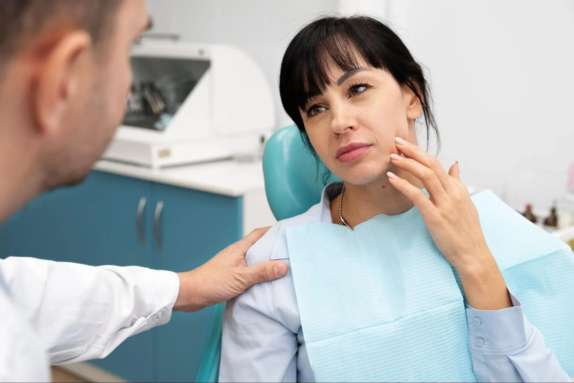
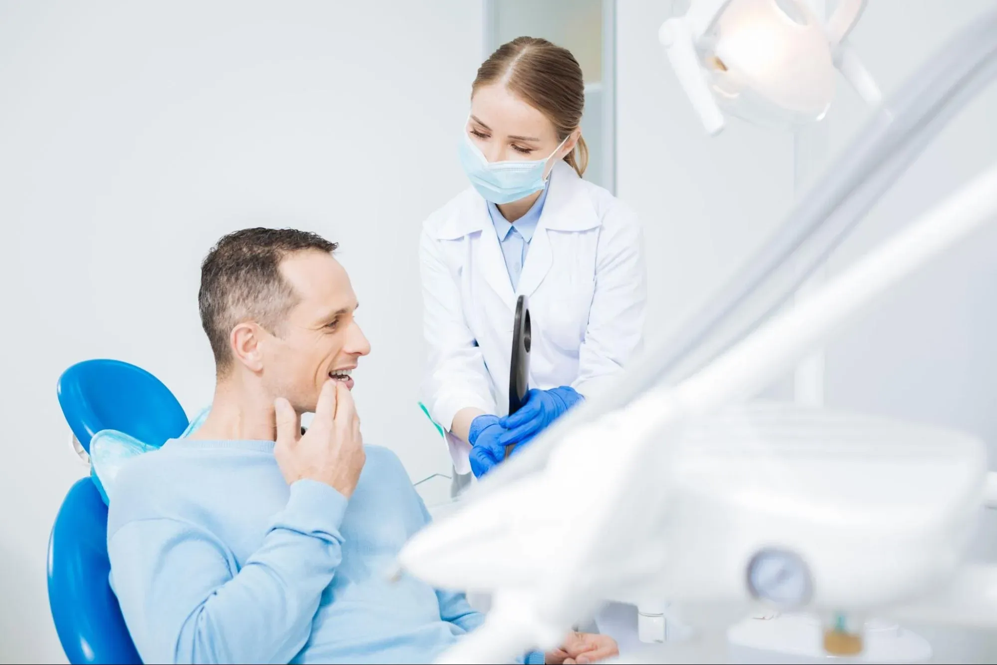
That's why the Department of Oral Diagnosis and Radiology is usually your first stop as a new patient. Before any treatment or cleaning, we need to understand what's happening beneath the surface.
Many patients feel nervous when they hear 'radiology' or 'X-ray,' often worrying about radiation. Some also wonder why a 3D scan is needed for a simple implant. This guide will explain the technology, the difference between 2D and 3D images, and the safety of modern digital diagnostics.
The Foundation: What is Oral Diagnosis?
Oral diagnosis means using both what we see in your mouth and what X-rays show to find problems and plan treatments. It's more than just looking for cavities. It includes:
-
Cancer Screening: Checking the soft tissues (tongue, cheeks, lips) for suspicious lesions.
-
TMJ Evaluation: Checking the jaw joint for clicking or wear.
-
Periodontal Assessment: Measuring gum pockets and bone levels.
-
Radiographic Analysis: Using X-rays to see inside the bone.
A good treatment plan depends on getting the diagnosis right. If the diagnosis is off, the treatment won't work.
The Evolution: Analog vs. Digital Radiology
In the past, dental X-rays meant putting a sharp film in your mouth, getting a quick burst of radiation, and waiting about 10 minutes for the film to develop in a room with chemicals. Now, we use digital radiography (RVG).
The Digital Advantage:
-
Low Radiation: Digital sensors are incredibly sensitive. They require 80-90% less radiation than traditional film to capture an image.
-
Instant Results: The image appears on the computer screen in seconds.
-
Enhanced Diagnostics: Dentists can zoom in, change the contrast, and highlight parts of the image to find small cracks or early decay that film might miss.
-
Eco-Friendly: No more toxic developing chemicals.
Types of 2D Imaging: The Standard Maps
Most dental check-ups use two-dimensional X-rays, which give a flat image of your teeth.
1. Periapical X-Ray:
-
The View: Focuses on just 2 or 3 teeth, showing the entire length from the crown to the root tip and the surrounding bone.
-
The Use: Essential for diagnosing root canal infections and abscesses, or for checking the fit of a restoration.
2. Bitewing X-Ray:
-
The View: Shows the crowns of the upper and lower back teeth biting together.
-
The Use: This is the best way to find 'interproximal decay,' or cavities that form between teeth where you should floss. These cavities can't be seen until they get very large.
3. Panoramic X-Ray (OPG):
-
The View: This is a wide-angle image that shows your whole mouth, including all your teeth, jaw joints (TMJ), and sinuses, all in one picture.
-
The Use: This is the usual scan for check-ups. It's great for looking at wisdom teeth, bone levels, children's development, and for checking for jaw cysts or tumors.
The Game Changer: 3D Dental Tomography (CBCT)
Panoramic X-rays are helpful, but they only show 2D images of 3D objects, so some things can be hidden. That's where Cone Beam Computed Tomography (CBCT) comes in. Unlike large medical CT scanners where you lie down, a dental CBCT is an open, upright machine that circles your head once in about 10 to 20 seconds.
Why is 3D necessary?
-
Implant Planning: To place a titanium screw in the jaw, we need to know the exact size of the bone and where the main nerve is, so we don't risk nerve damage. CBCT lets us measure this with 0.1mm accuracy.
-
Impacted Teeth: For wisdom teeth tangled around a nerve, a 3D scan shows the surgeon exactly which angle to approach, reducing surgical risk.
-
Endodontics: Sometimes a root canal fails because the tooth has a tiny 4th canal that is hidden behind the others on a 2D X-ray. CBCT reveals this hidden anatomy.
-
Pathology: CBCT helps us see the exact size and edges of a cyst or tumor before surgery.
The Elephant in the Room: Radiation Safety
"Is it safe?" This is the question we hear most often. We follow the ALARA Principle, which means we keep radiation as low as reasonably possible and only take X-rays when needed.
To put it in perspective:
-
We are exposed to "background radiation" every day from the sun, the soil, and cosmic rays.
-
A single digital dental X-ray gives you about the same amount of radiation as eating a few bananas or taking a two-hour flight.
-
A panoramic X-ray is equal to the radiation you get from one to three days of normal living.
-
A Dental CBCT varies, but modern machines use a "pulsed" beam to keep the dose very low compared to a hospital medical CT (which can be 50-100 times higher).
The risk from an untreated infection, like an abscess spreading to the heart or brain, is much higher than the very small risk from a modern dental X-ray.
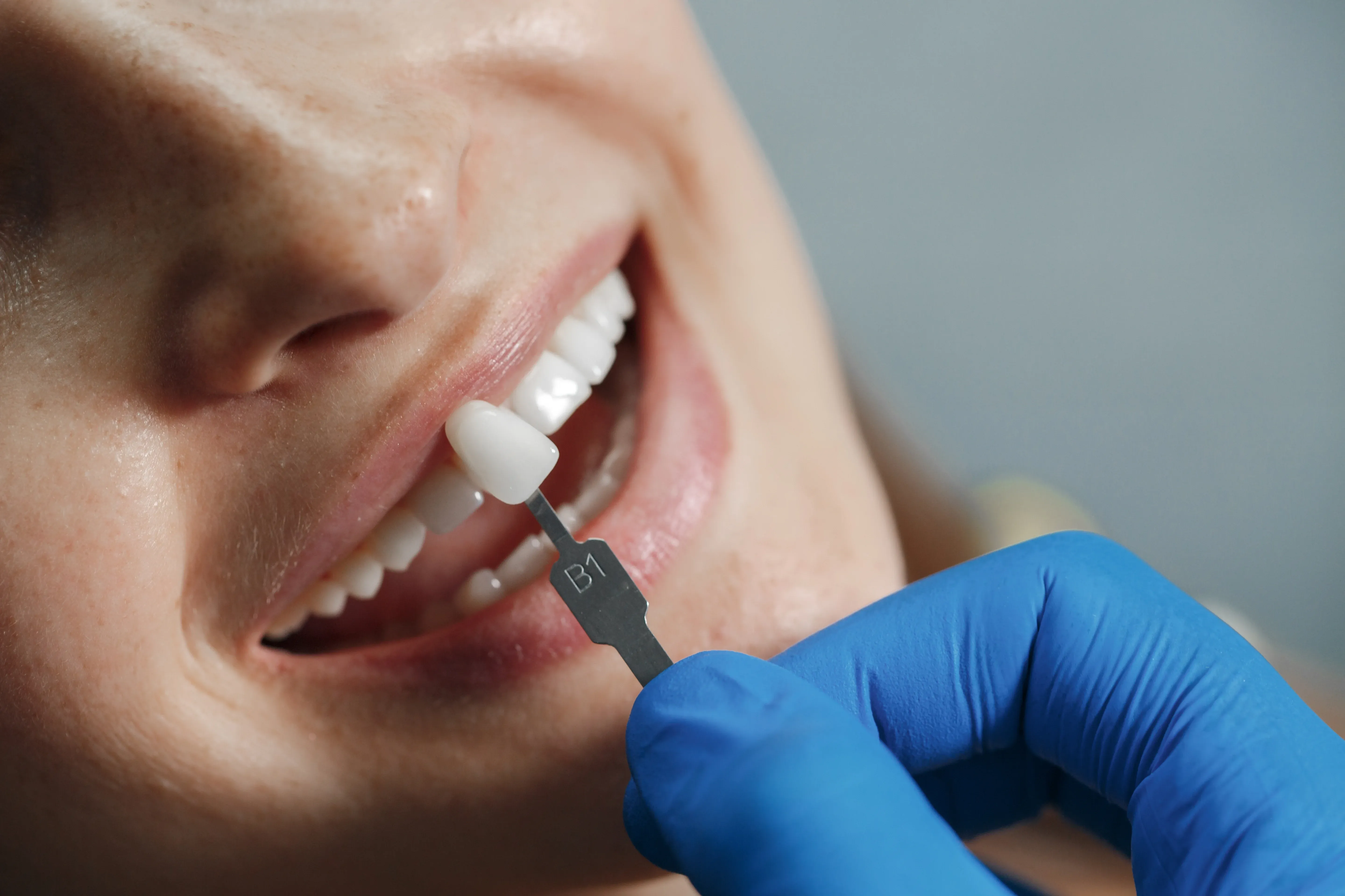
Beyond Radiation: Intraoral Cameras
Diagnostics involve more than just X-rays. Intraoral cameras are small, wand-shaped tools with high-definition video lenses.
-
The Benefit: We use the camera in your mouth and show you a live video on the screen. You can see cracks in old fillings, tartar behind your lower teeth, or a red spot on your gum.
-
Co-Diagnosis: This changes things from 'The dentist told me I have a cavity' to 'I saw the cavity myself.' It helps you understand your own dental health.
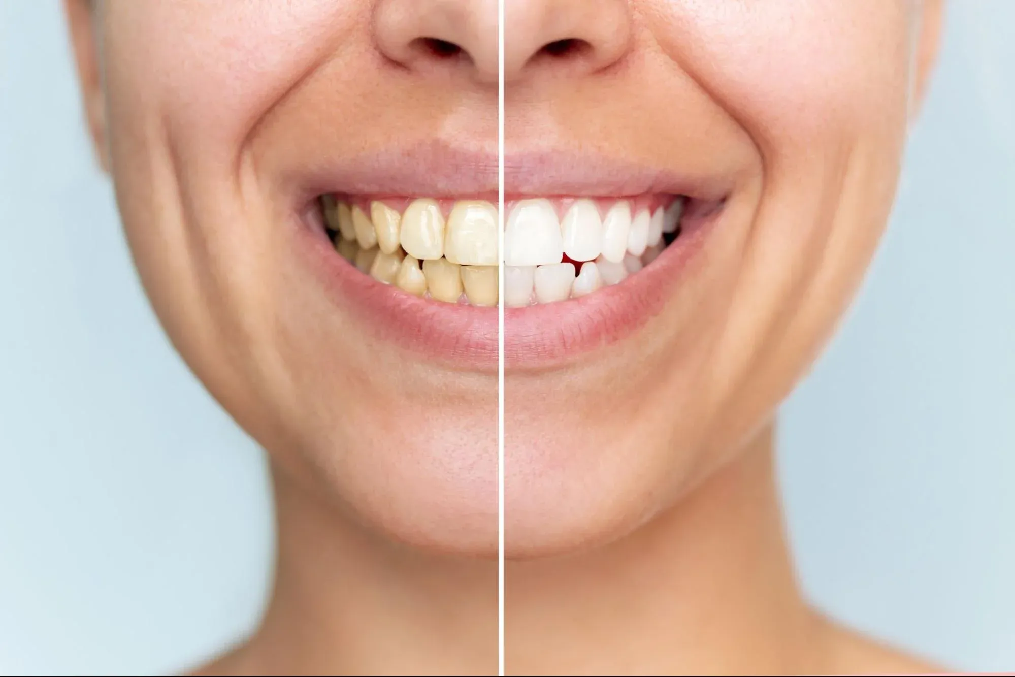
Early Diagnosis Saves Teeth (and Money)
The main goal of the Diagnostic Department is to catch problems early. Finding a cavity when it's just a small spot means a quick, inexpensive filling. Waiting until it hurts often means a root canal and crown. Finding bone loss early means a deep cleaning, but waiting until the tooth is loose means it may need to be removed and replaced.
Radiology helps us act early instead of waiting for problems to get worse.
The First Step of Your Journey
At İstinye University Dental Hospital, we see the diagnostic appointment as the most important visit. We use the latest low-dose digital panoramic and periapical sensors, as well as advanced CBCT units, for planning complex surgeries.
Our radiologists and oral diagnosis specialists are like the architects of your care. They collect information, look for hidden risks, and work with other specialists to create a treatment plan that is safe, accurate, and clear. In dentistry, just like in life, the best choices come from having the best information.